AI 生成イラスト
ログイン
AI 生成イラスト
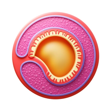
Anatomy
Gallbladder
Gallbladder
コミュニティ内で何も見つかりませんでした
生成結果